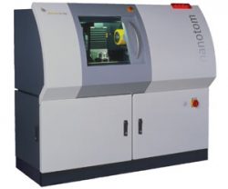
Description
Key Features
- First 180 kV / 15 W nanoCT® system with low maintenance, longlife open X-ray tube
- High precision rotation-unit with air bearing for accurate and reliable CT images
- Granite-based manipulation and optional thermal stabilization for long-term stability and highest precison
- Up to 2 times faster data acquisition at the same high image quality level by diamond|window (optionally)
Customer Benefits
- 4 operation modes from submicron to high-intensity applications
- Automated generation of first article inspection reports in < 1 hour possible
- Excellent software modules for highest CT quality and ease of use, e.g.
- High precision and reproducible 3D metrology by click & measure|CT with datos|x 2.0: fully automated execution of CT scan,reconstruction and analysis process
- Accelerated 3D CT reconstruction results within a few seconds or minutes (depending of the volume size) by velo|CT
- Very compact system even for small labs
Applications
3D Computed Tomography
The classic application of industrial X-ray 3D computed tomography (micro ct and nano ct) is the inspection and three-dimensional measurement of metal and plastic castings. However, phoenix|x-ray’s high-resolution X-ray technology opens up a variety of new applications in fields such as sensor technology, electronics, materials science, and many other natural sciences.
 nanoCT® of a SMD-inductor, size 0805 (2.0 mm x 1.2 mm). The 3D X-ray image shows the interior coil behind the end cap. In any conventional radiograph, the layers would be overlapping, but the nanoCT® succeeds in displaying the object layer by layer.
Material Science
High-resolution computed tomography (micro ct and nano ct) is used for inspecting materials, composites, sintered materials and ceramics but also to analyze geological or biological samples. Materials distribution, voids and cracks are visualized three-dimensionally at microscopic resolution.
 nanoCT® of a glass fiber-composite material: The fiber direction of the fiber mats (blue) and the matrix resin (orange) are displayed. Right: Voids inside the resin appear as dark cavities. Left: The resin has been faded out to better visualize the fiber mats. The individual fibers inside the mat are visible.
Sensorics and Electrical Engineering
In the inspection of sensors and electronic components, high-resolution X-ray technologies are mostly used to inspect and evaluate contacts, joints, cases, insulators and the situation of assembly. It is even possible to inspect semiconductor components and electronic devices (solder joints) without having to disassemble the device.
 nanoCT® showing solder joints of a CSP-component. The three-dimensional shape of the solder joints, app. 400 µm in diameter, and void distribution are clearly visible. Inside the solder joints, different eutectic solder phases are visible.
Metrology
3D metrology with X-ray is the only technique allowing to non-destructively measure the interior of complex objects. By contrast with conventional tactile coordinate measurement technique, a computed tomography scan of an object acquires all surface points simultaneously – including all hidden features like undercuts which are not accessible non-destructively using other methods of measurement. The v|tome|x s has a special 3D metrology package that contains everything needed for dimensional measuring with the greatest possible precision, reproducibility and user-friendliness, from calibration instruments to surface extraction modules. In addition to 2D wall thickness measurements, the CT volume data can be quickly and easily compared with CAD data, for example, in order to analyse the complete component to ensure it complies with all specified dimensions.
 CAD variance analysis and measurement of three features of a cylinder head.
Plastics Engineering
In plastics engineering, high-resolution X-ray technology is used to optimize the casting and spraying process by detecting contraction cavities, blisters, weld lines and cracks, and to analyze flaws. X-ray computed tomography (micro ct and nano ct) provides three-dimensional images of object characteristics such as grain-flow patterns and filler distribution as well as of low-contrast defects.
 nanoCT® of a sample of glass fiber-reinforced plastic: Alignment and distribution of the glass fibers and agglomerations of mineral filler (purple) are clearly visible. The fibers are app.10 µm wide.
Geology / Biological Sciences
High-resolution computed tomography (micro ct and nano ct) is widely used in inspecting geological samples, for example in the exploration for new resources. High-resolution CT-systems provide three-dimensional images at microscopic resolution of rock samples, binders, cements and cavities and help identify certain sample characteristics such as size and location of voids in oil-bearing rock
 Segmented pores in carbonate (ø 2 mm)
Specifications
| Max. tube voltage | 180 kV |
|---|---|
| Max. output | 15 W |
| Detail detectability | Up to 200nm (0.2µm) |
| Min. focus-detector-distance | 0.4mm |
| Max. voxel resolution (depending on object size) | < 500nm (0.5µm) |
| Geometric magnification (3D) | 1.5 times up to 100 times |
| Max. object size (height x diameter) | 150mm x 120mm / 5.9″ x 4.7″ |
| Max. object weight | 2 kg/ 4.4 lb |
| Image chain | 5-Megapixel fully digital image chain |
| 2D X-ray imaging | no |
| 3D computed tomography | yes |
| Advanced surface extraction | yes (optional) |
| CAD comparison + dimensional measurement | yes (optional) |
| System size | (1640 x 1430 x 750 mm), (64.6” x 56.3” x 29.5”), larger cabinets on request |
| System weight | 1300kg / 2866 lb |
| Radiation Safety | – Full protective radiation safety cabinet according to the German RöV (attachment 2 nr. 3) and the US Performance Standard 21 CFR 1020.40 (Cabinet X-ray Systems) – Exposure rate < 1 µSv/h emission limit measured at 10 cm distance from accessible surfaces |
Download

